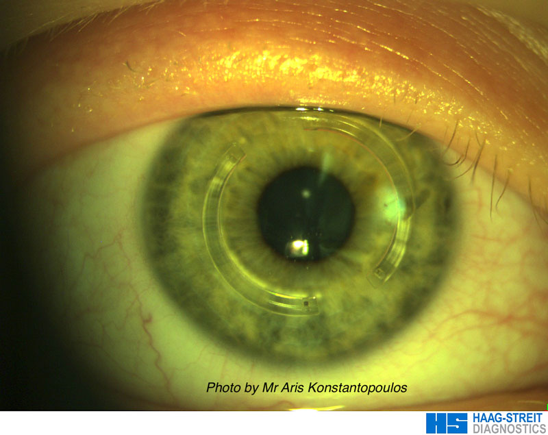
What is keratoconus?

Keratoconus is a slowly progressive condition in which the cornea, the clear round window at the front of eye, becomes weaker and bulges forward. The cornea gradually changes shape from a sphere (like a football) to a cone (like a rugby ball), resulting in distorted and reduced vision.

Symptoms

The change in shape alters the refractive properties of the cornea, reducing the quality of vision. A key feature of the condition is a gradual progression in myopia (short-sightedness) and astigmatism.

As the condition becomes more advanced and the astigmatism irregular, spectacles fail to compensate for this, resulting in blurred vision. Other symptoms of keratoconus can be distorted vision, double or multiple images, and streaks or haloes around lights.

Cause

The cause of keratoconus is not fully understood but the condition is considered to develop due to a combination of genetic and environmental factors. However, it is not thought of as an inherited condition. There is a good association with atopy and often patients also have allergic eye disease with itchiness and rubbing of the eyes.

How is keratoconus diagnosed?

Detection by your optician of rapidly progressive myopia and astigmatism can be a sign of keratoconus and requires further investigations. These investigations are usually carried out by an ophthalmologist (eye doctor) who will carry out a detailed examination with a slit-lamp biomicroscope and corneal topography imaging. Corneal topography involves detailed scanning of the cornea, plotting of its shape and comparison to the expected normal.

How can keratoconus be treated?

Importance of early diagnosis

Early diagnosis of the condition is key to maintaining good vision. In the early stages, the vision can be improved with glasses or soft contact lenses. Treatment at this stage will maintain the best possible vision for the long term.

As the condition progresses and the cone become more advanced, the astigmatism becomes irregular and rigid gas permeable (hard) contact lenses are required to improve the vision.

As the condition tends to be progressive in nature, wearing glasses and contact lenses does not stop the underlying progression.

Collagen cross-linking

Corneal collagen cross-linking is the only proven treatment that can stop progression of the condition; however, it does not reverse changes in the shape of the cornea or any visual loss that may have already developed. In September 2013, the National Institute of Clinical Excellence (NICE) recommended the use of epithelium-off collagen cross-linking for keratoconus and keratectasia (Interventional procedures guidance IPG466).

In collagen cross-linking, riboflavin (B2) eye drops interact with UVA light to stimulate the formation of extra bonds (cross-links) between the collagen fibres of the cornea, thus increasing its rigidity and strength.

Collagen cross-linking can be combined with laser vision correction; this combined procedure not only stops progression of the condition but can also improve the quality of your vision, especially in the early to moderate stages.

Corneal-ring-patient

Laser treatment

Topography guided advanced surface ablation can improve the vision in patients with early to moderate keratoconus. The excimer laser treatment is customised to the shape of the cornea, resculpting the corneal surface, and making the cornea more like a sphere. This reduces the irregular astigmatism and aberrations, improving the quality of vision.

Laser treatment can be performed after corneal collagen cross-linking has been carried out or the two procedures can be combined. The combination of the 2 procedures improves the strength of the cornea and also the quality of vision. The combined treatment is often referred to as the Athens protocol for keratoconus.

Intrastromal corneal ring segments

Intrastromal corneal ring segments are small circular implants that are placed within the cornea. Their placement is guided by the precise creation of corneal channels with the use of our Visumax femtosecond laser.

Their size and positioning are matched to the shape of your cornea, flattening the steep areas and returning the conical cornea to a more normal shape. This results in a reduction in myopia (short-sightedness), astigmatism and aberrations, improving your quality of vision.

Corneal transplantation

In advanced cases of keratoconus, in which the above methods of visual correction are not applicable, the vision can be improved with corneal transplantation. With the increasing awareness of keratoconus and its earlier detection, the requirement for transplantation should decrease in the future.

In corneal transplantation the ectatic and weaker cornea is replaced by a healthy donor cornea, restoring the normal optical properties of the eye and improving vision. Transplantation can take the form of a full thickness corneal graft, known as penetrating keratoplasty, or a partial thickness corneal transplant, known as deep anterior lamellar keratoplasty (DALK).